Object ID Number:
EH101379
Object Name:
Electrocardiograph Machine
Type:
EKG Machine; Sanborn Cardiette
Manufacturer:
Sanborn, Cambridge, MA
Date of Manufacture:
/ /
Manufactured from:
1925
Manufactured to:
1945
Description / History:
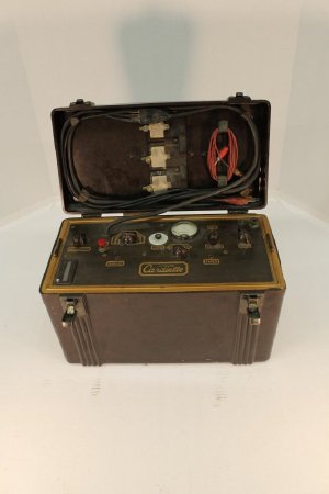
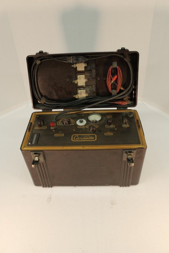
This EKG machine, also referred to as the Sanborn Cardiette, was manufactured by Sanborn in Cambridge, MA. The casing is made from bakelite, an early plastic, and brown in color. The case comes with a handle to make the EKG portable when necessary. Inside the case there are various cords, metal pads, a meter, and several dials (reading: "Sensitivity", "IMV". "Leads Batteries", "Marker" , "Lamp", "On Camera Off" , " Voltage" and "Beam"). On the side of the machine there is a removable door where the film is stored to record the electric currents. On the back of the machine there is a crank that can be spun. A very similar machine with the same type of cords and same case style except for wood used as material is located at the Collection of Science Instruments at Harvard University and dates to about 1925. Since plastic started to be used on occasion in the 1930s, we can concluded it is around this time frame. The Sanborn Cardiette was used only for a little while until Sanborn invented the Sanborn Viso Cardiette in 1946 which allowed the doctor to visually watch the currents being recorded through a glass screen and then printed for their convenience.
Dimensions:
H–9.25 W–7 L–15 inches
Additional Information:
Electrocardiograms (EKGs)
An electrocardiogram (also called ECG or EKG) is a noninvasive test that records the electrical activity of the heart. This examination is useful because, with each heartbeat, an electrical signal spreads from the top to the bottom of the heart. As this electrical signal travels, it induces the heart to contract, and to pump blood throughout the body. Since the heart's electrical signals determine the rhythm of the heartbeat, an EKG can be used to show how quickly the heart is beating, whether the heart's rhythm is steady or irregular, and how strongly the electrical signals pass through each part of the heart. Based on these readings, a physician may discover a number of conditions, such as arrhythmias or recent heart attacks, and may also monitor the functioning of implanted pacemakers.
The earliest electrocardiogram was designed in 1901 by William Einthoven, a scientist from the Netherlands. In Einthoven's design, currents from the heart's electrical signals were carried down a silver coated glass conducting wire which was suspended between two electromagnets. The fluctuations of the wire as a result of the changing electrical currents were transcribed onto a photographic plate and produced readouts similar to those given by modern EKGs (although Einthoven's machine weighed 600 pounds, took up two rooms, and needed five people to operate).
EKGs remain one of the most important diagnostic tools today, and in 2011, 82,707 EKGs were performed at Lancaster General Hospital (or affiliated healthcare institutions), which amounts to a staggering 226 per day.
Click to Enlarge